
Cartilage Treatments
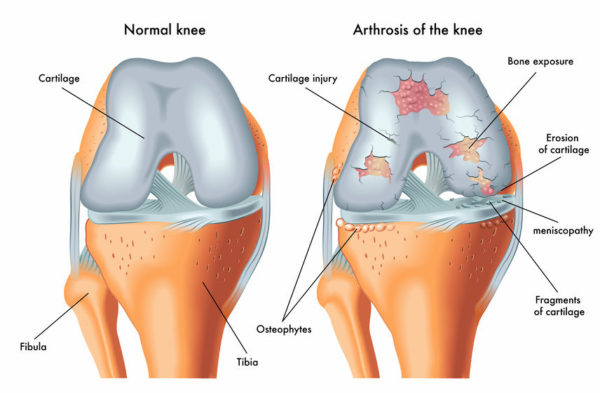 Where two bones meet within a joint, their surfaces are covered in protective articular cartilage which reduces the friction as bones glide over each other during movement and acts as a “shock absorber”. When this important joint surface layer is damaged, your surgeon may recommend surgery to help relieve the symptoms. Often, the first step in cartilage surgery involves gently removing (debriding) the damaged tissue.Unfortunately, while removing the damaged tissue can help to improve your symptoms for a time, debridement alone is ofteninsufficient to restoreyour joint surface permanently. This is because the lack of blood vessels in the cartilage layer means your body cannot get enough nutrients to the injured area, which makes rebuilding the protective surface difficult. However,new research and technology is making it possible to help stimulate healing in the affected areas and your surgeon offers several procedures using these techniques to treat cartilage injuries.
Osteochondral Repair
If a piece of cartilage shears away from an injured area, it will not heal back down to the joint surface as the cartilage layer does not have enough blood vessels to provide the nutrients needed for repair. The floating fragment is referred to as a loose body which can catch inside the joint with movement and cause pain or locking of your knee, as well as potentially causing more damage to your joint surface. It is often recommended that the loose cartilage be removed from the joint to relieve symptoms and prevent further joint surface damage.
However, when a piece of cartilage and the underlying (subchondral) bone breaks away, it leaves an osteochondral defect in the joint surface, along with the loose body inside the knee joint. Bone has a much better blood supply and is able to heal more readily than cartilage so in some cases, your surgeon may be able to attach the bony fragment with its overlying cartilage cap,back to the osteochondral defectin the joint surface (bone to bone). Once the loose fragment is replaced, it is usually anchored in place using specially designed screws or pins. This is known as osteochondral repair and is most oftenachieved using arthroscopic (key-hole) surgery. After this procedure, the replaced fragment will need time to heal so you may be advised to use crutches and practice a program of exercises with your physiotherapist to protect your knee as it recovers.
Microfracture
This technique might be recommended where the articular cartilage layer only has a small defect, surrounded by healthy joint surface. The cartilage layer does not have a good supply of blood vessels to provide the nourishment required for tissue healing. However, the subchondral bone that lies directly beneath it does have a good blood supply. The microfracturetechnique is an arthroscopic (key-hole) procedure which involves your surgeon creating tiny tunnels through the damaged cartilage layer, into the underlying bone. This allows cartilage forming “stem cells”and nutrientsto flow from the underlying bone, through these tunnels to restore the joint surface. These special cells are not able to perfectly replicate normal articular cartilage but instead, create a sort of cartilage “scar” which is able to function in a similar way to normal cartilage to help relieve pain and restore function to your knee.This rebuilding process is delicate so following this surgery, you will need to use crutches to reduce the pressure on this layer and work with a physiotherapist to ensure you get the most benefit from this procedure.
OATS procedure
Your surgeon may recommend this procedure for larger areas of cartilage damage inside your knee. Osteochondral autograft transfer system (OATS) involves carefully removing a cylinder shaped area of cartilage and underlying bone from your knee that includes the damaged part of joint surface. Then, a small segment of healthy bone and cartilage (autograft) is harvested from an area of your own knee which does not carry weight when you move. This healthy graft is the same size and shape as the area of damaged tissue that has been harvested from your knee so that when the autograft cylinder is placed into the defect, it is a perfect fit and leaves a smooth and continuous joint surface. It is possible to repeat this procedure with several smaller graft plugs to create a “mosaic” of existing and transplanted joint surface. For this reason, this procedure is known as a mosaicplasty. Following either OATS or mosaicplasty, the transplanted bone and cartilage will need time to integrate into the joint surface so it is important to use crutches after the procedure and to work with your physiotherapist to give your knee the best chance of healing.
Cartilage Restoration
Some exciting new technologies are being developed to help grow new articular cartilage to repair areas of joint surface that have been damaged. An example of this is the use of bioscaffolds which are highly specialised products, designed to be placed inside a defect in the joint surface to provide a nourishing and protective platform into which new cartilage can develop. These products are eventually broken down by the body in a harmless process, leaving behind the rebuilt joint surface. Scaffolds can be used in conjunction with other cartilage repair procedures, such as microfracture, to provide a stable environment for the stem cells once they flow from the subchondral bone to the joint surface. They can also be combined with other products and techniques that help to multiply the stem cells found in our own bodies which have the capacity to form new, healthy cartilage.
This area of medicine is always expanding. Your surgeon is familiar with these technological advances and can discuss these options with you so that you can decide together, which option might best suit you.